research supporting
the use of silicone in medical devices had been compiled before Cronin and
Gerow employed it in the mammary prosthesis.
1962 marked the first implanting of a mammary prototype. For the next two
years, selected surgeons used the implants in clinical trials to obtain
information on their performance, both long- and short-term, before Dow Corning
took the Cronin implant fully to market in 1964. Additional support for the
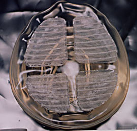
implants was found in the hydrocephalic shunt's performance results, since it
used the same elastomer. By 1962, four-thousand shunts had been placed in
children's brains, treating their hydrocephalus and saving their lives, all
without any apparent adverse effects [36].
Also, in 1962, the National Institute of Health funded the Battelle Memorial
Institute to conduct research[37] on the
stability of silicone implants in animals. The Institute's two-year study
focused on the effect of the body on the various polymers studied, which
included Polyethylene, Teflon, Mylar, Nylon, and Silasticreg.. In particular,
the study concentrated on the polymers' tensile strength and elongation, along
with the reaction of the implant site to the polymers. (Tensile strength is a
measure of the polymer's ability to withstand elongation forces.) The studies
used mongrel dogs as test sites, implanting samples of all five materials in
each. The plastics were recovered after six, eleven, and seventeen month
intervals. Their tensile strength was recorded before implantation and after
each removal, to track any loss, along with any elongation due to implantation.
After 17 months, Silasticreg. showed little decrease in tensile strength and
slight elongation. According to the study:
Changes [concerning tensile strength and elongation] in the Silasticreg.
and Mylar are not believed significant. . . In general, minimal local tissue
reaction was observed grossly at nacropsy. There was no gross evidence of
tumorigenesis either at the site of implant or in other organs and tissues.
No irritation or inflammation was seen . . . [However, the Silasticreg.
implant was observed to have] a poorly defined capsule which was
accidentally incised. As in previous necropsies, a great deal of difficulty
was encountered in locating the site of the flank implant. The Silasticreg.
strips were folded and wadded into a ball - no evidence of irritation or
local tissue reaction other than the connective tissue encapsulation. . .[38]
(Encapsulation is a common, but unharmful, foreign body reaction to any
implanted device, where tissue walls the device off from the rest of the body[39].) The
study also provided a histological
report on the 17-month findings. (Histology is the study of tissues[40].) The histological findings on
Silasticreg. showed "fibrous tissue segment without apparent inflammatory
reaction."
Simultaneous to the introduction of the Cronin implant to market in 1964, Dow
Corning contracted Food and Drug Research Laboratories, an independent research
company, to complete more long-term testing on the implants, which had already
been followed for two years in the clinical studies and seventeen months in the
Battelle study. In the three-year Food and Drug Research Laboratories
experiment, eleven polysiloxane compositions were implanted ". . . into
intramuscular, subcutaneous, and intraperitoneal sites in sufficient numbers to
permit samples and surrounding tissue to be removed at a number of periods
prior to sacrifice of the animals . . . The design of the study provided for
replication of each treatment in three dogs, restricting one type of
polysiloxane within each given dog, but permitting as many as four
replications of each product and site combination in a given animal."[41]. These silicones were implanted "over
protracted periods" in five forms: "solid film, perforated film, sponge,
amorphous forms, and miniature artificial breasts."[42] Microscopic examination of tissue
reaction was completed at three, nine, twenty-four, and thirty-six months. The
results of the study, published in 1968, proved encouraging for the use of
silicone in medical implants, especially in mammary prostheses .[43]
Although implants were first targeted at mastectomy patients, even Cronin and
Gerow would have been able to surmise the general population's desire to use
the mammary prostheses for enhancement as well. Thus, other manufacturers
developed similar implants, in response to a market which grew as women opted
for cosmetic breast procedures. However, Dow Corning, where the implant
originated, remained the industry leader. Dow Corning continued to look for
improvements to the implant, developing a new outer lining in 1968. This
elastomer envelope, while thinner, was also seamless. (At times, the line
where the two halves of the original implant were joined was noticeable under
the upper portion of the breast.) The seamless envelope was all one elastomer,
a thinner covering that provided even more aesthetically pleasing results.
Go on to Part 6
 With over 10 years of research
already completed on it, silicone was a natural candidate for use in breast
reconstruction. In addition, it had already been utilized in other medical
applications, such as the hydrocephalic shunt and life-saving pace-maker
described earlier. These cases could provide information on the long-term
effects of silicone on the host.
With over 10 years of research
already completed on it, silicone was a natural candidate for use in breast
reconstruction. In addition, it had already been utilized in other medical
applications, such as the hydrocephalic shunt and life-saving pace-maker
described earlier. These cases could provide information on the long-term
effects of silicone on the host.